How could we miss?!
Paunovska et al.


Introduction
As you may already be aware, our preprint was recently released as a pre-proof on Elsevier. It has been a long journey, and The Offsc℞ipt Pharmacist, Stephanie Seneff, and I are immensely proud of this paper. It proposes a paradigm shift and highlights why this is necessary for multi-particle colloidal nanomedicine.1
However, we are currently facing a bit of a dilemma with no ideal solution: Maria stumbled upon this 2020 paper by Paunovska et al.2 just now. The issue: our own paper is already in the production phase, and we expect to receive our galley proofs in the coming days. There is simply no longer an option to integrate this study–even though it fully supports and indirectly validates our hypothesis.
Why is this paper so important?
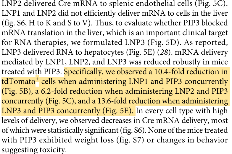
Paunovska et al. sought to understand why certain cells translate lipid nanoparticles with high efficiency while others do not. To test this, they experimentally elevated intracellular PI(3,4,5)P3 levels and systematically investigated the subsequent effects on LNP-mediated mRNA expression. They began by ruling out the most obvious explanations: neither cell viability, nanoparticle uptake, nor endosomal escape could account for the observed effect. Instead, they discovered that elevated PI(3,4,5)P3 levels massively reduced functional mRNA translation both in vitro and in vivo, by approximately 6–almost 14-fold, depending on the LNP system used.
To elucidate these findings, they subsequently performed RNA sequencing and metabolomic analyses. Both datasets revealed that extensive shifts in the cellular state occurred just a few hours after PI(3,4,5)P3 administration. Transcriptomic analysis identified numerous differentially expressed genes and affected signaling pathways. In parallel, metabolomics showed a profound metabolic reprogramming, characterized by increased glycolysis, alterations in the pentose phosphate pathway, enhanced lipid and nucleotide synthesis, as well as indications of epigenetic methylation processes. After 24 hours, additional signs of a catabolic metabolic state emerged, including depleted amino acid levels and markers of oxidative stress. PCA and hierarchical cluster analyses clearly separated PI(3,4,5)P3-treated cells from untreated controls, illustrating that PI(3,4,5)P3 did not trigger an isolated molecular effect, but rather remodeled the entire cellular state.
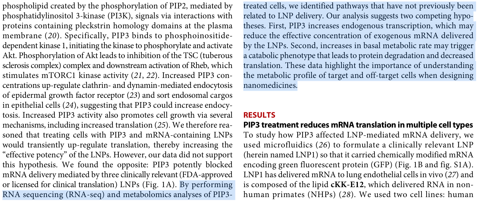
Ultimately, the authors interpreted these results to suggest that the elevated PI(3,4,5)P3 levels either consume cellular resources so heavily that exogenously introduced mRNA is essentially “drowned out,” or that the accelerated metabolism secondarily transitions into a catabolic state that impairs translation. Throughout their study, they repeatedly emphasize that translational efficiency normalizes after about 24 hours, and that the observed effect specifically relates to the cells’ ability to efficiently express therapeutic mRNA. Their conclusion, therefore, is that metabolic signaling pathways regulate the efficiency of LNP-mediated RNA expression.
However, this is precisely where our hypothesis comes in and goes significantly further.
This is because Paunovska et al. exclusively investigate the translational efficiency of the introduced mRNA. They do not question whether the cell returns to its original biophysical state after 24 hours. Similarly, they examine neither the long-term organization of the membrane, the dynamics of phosphoinositide pools, nor the restoration of the supramolecular membrane architecture. In other words, the observed “normalization” pertains solely to the functional endpoint of mRNA expression, rather than the underlying membrane state.
For our hypothesis, however, this exact underlying state is critical. We postulate that the primary disruption does not lie within a single signaling pathway, but in the physicochemical organization of the membrane itself. Phosphatidylinositides do not function merely as signaling molecules; they act as central organizers of membrane platforms. If this organization is disrupted, far-reaching alterations across numerous signaling networks inevitably occur simultaneously. From this perspective, the transcriptomic and metabolomic changes observed by Paunovska appear not as the cause of reduced translation, but as secondary, systemic consequences of a disrupted membrane organization.
Notably, despite unchanged nanoparticle uptake and no signs of relevant toxicity, Paunovska documents massive alterations in the overall cellular state. This aligns perfectly with the predictions of our hypothesis: a simple shift in phosphoinositide dynamics is sufficient to alter the spatial organization of membrane-associated processes, thereby simultaneously impacting the transcriptome, metabolism, and ultimately, translation. The fact that the authors themselves interpret this finding as a metabolic phenomenon does not contradict our hypothesis. Rather, it describes a potential downstream layer of the very same process.
From our perspective, the true strength of this study does not lie in the assertion that PI(3,4,5)P3 inhibits mRNA translation. Instead, the critical takeaway is that Paunovska experimentally demonstrates just how sensitively LNP function responds to comparatively minor changes in the phosphoinositide system. Consequently, the study provides direct experimental evidence that phosphoinositide homeostasis and LNP function are tightly coupled. This exact coupling forms the mechanistic core of our L-DMD hypothesis.
While Paunovska describes the pathway of PI(3,4,5,)P3→altered cell physiology→reduced translation, our hypothesis expands this model by addressing the upstream question of whether LNPs themselves, via their ionizable lipids, can disrupt phosphoinositide organization and thus the physicochemical self-organization of biological membranes. In this sense, the work of Paunovska is not a counterargument to our hypothesis, but rather one of its strongest experimental pillars, demonstrating that disruptions in phosphoinositide homeostasis alone are sufficient to trigger comprehensive cellular reprogramming.
Another crucial aspect is that Paunovska et al. identify phosphoinositide metabolism not as a passive background factor, but as a limiting regulator of LNP function. In their experimental design, the nanoparticle remains unaltered; only the cellular phosphoinositide state is manipulated. Yet, the efficiency of the entire LNP-mediated gene expression changes drastically. This clearly demonstrates that the functional success of an LNP is not determined solely by its chemical composition, but depends heavily on the membrane-physiological state of the target cell: a reciprocity that lies at the very core of our hypothesis.
Furthermore, their study underscores that phosphoinositides are not isolated signaling lipids, but master regulators of cellular organization. The observed alterations are not confined to a single pathway; they encompass the transcriptome, the metabolome, and ultimately, the cell’s capacity to efficiently express exogenous mRNA. This pattern points to a system-wide reorganization rather than a localized molecular event. From the perspective of our hypothesis, this is precisely the behavior to be expected when the organization of membrane-associated signaling platforms is disrupted.
The direction of causality here is of particular importance. While Paunovska et al. demonstrate that experimentally altering the phosphoinositide system is sufficient to trigger profound functional consequences, our hypothesis poses the complementary question: Can ionizable lipids themselves initiate such a disruption in phosphoinositide homeostasis? If so, the molecular and metabolic shifts described by Paunovska would not be independent phenomena, but rather the predictable consequences of a primary, membrane-physical disruption.
The study also carries significant methodological weight. It reveals that evaluating LNPs based solely on classic endpoints–such as cell viability, nanoparticle uptake, or endosomal escape–can be inadequate. Despite unchanged uptake and a lack of overt cytotoxicity, fundamental cellular programs are altered. Thus, the study indirectly supports our premise that functional changes in biological membranes can occur without manifesting in conventional toxicity parameters. This directly aligns with our argument that regulatory safety assessments focusing primarily on acute immune responses or cell damage may overlook potentially critical membrane-biophysical effects.
Ultimately, Paunovska et al. illustrate that phosphoinositides are far more than downstream effectors of individual signaling cascades; they establish the functional prerequisites for cellular processes to occur efficiently in the first place. This paradigm shift is central to our hypothesis: phosphoinositides are understood not merely as another component within a pathway, but as organizing elements of the membrane architecture itself. This shifts the focus from the regulation of isolated pathways to the integrity of the physicochemical platform upon which these networks are assembled and coordinated.
It is precisely for this reason that this paper is so vital to our hypothesis. It provides definitive experimental proof that targeted alterations in phosphoinositide homeostasis suffice to induce far-reaching functional changes across multiple biological tiers. Our work expands upon this finding by proposing a mechanism: that ionizable lipids from LNPs can themselves disrupt phosphoinositide organization, thereby serving as the common denominator for the wide array of observed molecular and cellular changes. This positions their study as one of the most compelling independent experimental pillars supporting the L-DMD hypothesis.
The elephant in the room
Despite its experimental elegance, the conceptual framework adopted by Paunovska et al. remains largely confined to the traditional molecular and cellular biology paradigm. Phosphoinositides are primarily interpreted as signaling lipids whose altered abundance modulates downstream metabolic and transcriptional pathways. Within this framework, PI(3,4,5)P₃ is effectively treated as an independent experimental variable whose increase perturbs cellular physiology.
However, biological membranes are not collections of isolated signaling molecules. PI(4,5)P₂ and PI(3,4,5)P₃ exist as integral components of a highly dynamic phosphoinositide network, in which local synthesis, turnover, lateral diffusion, and membrane compartmentalization are continuously coordinated in both space and time. Consequently, the biological significance of phosphoinositides cannot be reduced to their absolute abundance alone; rather, it emerges from their spatial distribution, local stoichiometry, and dynamic organization within the membrane.
This raises a fundamental question that remains unexplored. How did experimental elevation of PI(3,4,5)P₃ alter the local PI(4,5)P₂/PI(3,4,5)P₃ balance at the cytoplasmic membrane leaflet? Did these changes modify lipid packing density, membrane curvature, nanoscale lipid domain formation, or the clustering and lateral mobility of membrane receptors? Equally important, how were the electrostatic properties of the inner membrane leaflet affected, and what consequences did this have for the recruitment and dissociation of phosphoinositide-binding proteins that orchestrate membrane trafficking and signal transduction?
These questions are not merely technical details but address a fundamentally different level of biological organization. Phosphoinositides are not simply signaling intermediates; they are key organizers of membrane architecture. Through their unique electrostatic properties, they govern the spatial recruitment of peripheral membrane proteins, define membrane identity, and establish the physicochemical environment in which signaling complexes assemble and function.
From the perspective of the L-DMD hypothesis, this distinction is critical. The widespread alterations in the transcriptome, metabolome, and translational capacity observed by Paunovska et al. are unlikely to represent the initiating event. Rather, they are more plausibly interpreted as downstream manifestations of a preceding disruption in the spatiotemporal self-organization of the phosphoinositide network and, consequently, of the biological membrane itself. In this view, membrane organization is not simply the substrate upon which signaling occurs: it is the primary level at which dysregulation is initiated.
Conclusion
Paunovska et al. convincingly demonstrate how sensitive cellular physiology is to perturbations of the phosphoinositide system. What remains unresolved, however, is the more fundamental question of how the physicochemical organization of this system itself is altered. Likewise, the systemic consequences of large-scale membrane perturbations induced by lipid nanoparticles remain largely unexplored. If the L-DMD hypothesis is correct, such perturbations would be expected to propagate far beyond isolated signaling pathways and could result in highly heterogeneous biological outcomes. The critical questions are therefore no longer whether membrane organization matters, but rather to what extent its disruption contributes to long-term biological consequences, under which conditions, and in which tissues.

However, the question remains: How could we just miss this work?!
Seger, F., Gutschi, L.M. & Seneff, S. (2026) ‘Lipid nanoparticles as active biointerfaces: From membrane interaction to systemic dysregulation’, Acta Pharmaceutica Sinica B. doi:10.1016/j.apsb.2026.07.001
Paunovska, K., Da Silva Sanchez, A., Foster, M.T., Loughrey, D., Blanchard, E.L., Islam, F.Z., Gan, Z., Mantalaris, A., Santangelo, P.J. & Dahlman, J.E. (2020) 'Increased PIP3 activity blocks nanoparticle mRNA delivery', Science Advances, 6(30), eaba5672. doi:10.1126/sciadv.aba5672.
 | Ein Gastpost von
|
https://tkp.at/2026/07/13/wie-lipid-nanopartikel-aus-den-mrna-spritzen-aktiv-in-die-zellarchitektur-eingreifen/ Congrats!