Growing evidence
LDMD–An Inconvenient Hypothesis

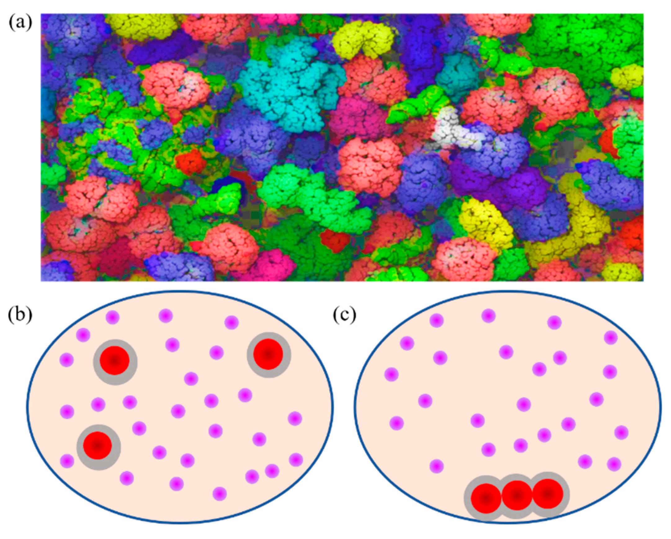
***1
This brief article discusses two recent studies that provide new insights into LNP-mediated delivery and the role of biological context. Our hypothesis2 is receiving increasing support from emerging evidence that LNP performance is governed not only by nanoparticle composition but also by the biological context in which LNP–cell interactions occur.
A recent study published by Kunitskaya et al.3 partially confirms our physicochemical considerations that the key factor is not solely the optimization of the LNP formulation itself.
The study demonstrated that increasing T-cell concentration during transfection increased the number of transfected cells per ng of mRNA by up to 30-fold, while the overall transfection rate remained unchanged at approximately 55%. At a cell concentration of 30 × 106 cells/mL, the authors achieved approximately 55% GFP-positive T cells (i.e., essentially the same transfection rate as in the standard protocol) while requiring approximately 40-fold less mRNA. Consequently, the number of transfected cells per ng of mRNA used increased by approximately 30-fold.
The authors attribute this effect to the increased probability of LNP–cell interactions at higher cell densities, leading to enhanced LNP binding and uptake rather than allowing LNPs to remain dispersed within the suspension. Washing experiments further supported this hypothesis. Importantly, the benefit of increased cell concentration was observed independently of the specific LNP formulation used and improved transfection efficiency by approximately 20-fold on average.
Although NK cells are generally more challenging to transfect, the method also increased transfection efficiency in NK cells by approximately 10-fold per ng of mRNA. Notably, only 1 hour of incubation at room temperature was sufficient to achieve this effect. Another interesting observation by the authors was that room temperature performed at least as well as 37°C and, at very high cell concentrations, even provided advantages in terms of cell viability and expansion.
Further increasing the cell concentration to 100 × 10⁶ cells/mL improved mRNA utilization even further; however, this came at the expense of reduced cell viability. Therefore, 30 × 106 cells/mL currently appears to represent the most practical compromise.
The maximum transfection rate in primary T cells remained approximately 50–55%. The authors suggest that further improvements will likely require optimization of the LNP composition, particularly the ionizable lipid component. In addition, substantial batch-to-batch variability of the LNPs was observed.
Importantly, the study primarily evaluated functional transfection outcomes based on eGFP expression, flow cytometric analysis of GFP-positive cells, cell viability measurements, and selected cytokine-related readouts. It did not investigate potential density-dependent changes in LNP composition, protein corona formation, extracellular organization, particle behavior, or other physicochemical characteristics of the LNP–cell interface. Therefore, while the observed improvement clearly demonstrates a strong density-dependent enhancement of mRNA delivery, the underlying mechanism remains open to further investigation.
The interpretation that increased cell density primarily enhances transfection by increasing the probability of LNP–cell encounters may represent only the most immediate and measurable aspect of a broader density-dependent phenomenon. In the context of T cells, high-density conditions are known to generate emergent biophysical states associated with macromolecular crowding and collective cellular behavior.4,5, 6 This may also provide a potential explanation for the comparatively weaker effect observed in NK cells, which differ from T cells in their biology, surface organization, and sensitivity to density-dependent cell–cell interactions7.
Therefore, increased cell concentration may not merely increase the number of available cellular targets, but may also modify the physicochemical microenvironment in which LNP–cell interactions occur. It may lead not only to steric effects, such as altered diffusion behavior, molecular accessibility, and encounter kinetics, but also to electrochemical consequences, including changes in local ion distribution, interfacial charge environments, and hydration states within the extracellular space surrounding the LNP–cell interaction site.
This perspective would also align more closely with the concept proposed by us and by Trollmann et al.8: that LNP activity is governed not solely by the intrinsic properties of the nanoparticle, but by a dynamic biological interface comprising the LNP surface, the evolving protein corona, the surrounding extracellular microenvironment, and the interacting cellular membrane. In this view, LNP performance emerges from the interplay between nanoparticle physicochemistry and the local biological context in which particle recognition, membrane interaction, and intracellular delivery take place.
Interestingly, Trollmann et al. performed atomistic constant-pH molecular dynamics simulations to investigate how membrane environments regulate the protonation behavior of ionizable lipids. Their simulations included the following five widely used aminolipids:
DODAP
DLin-MC3-DMA
DLin-KC2-DMA
ALC-0315
SM-102
Importantly, the study’s central comparison focuses on the difference between the behavior of these lipids in isolation under infinite dilution conditions, where their intrinsic pKa values are defined and used for simulation parameterization, and their behavior when embedded within LNP-relevant membrane environments consisting of:
DOPC
DSPC
Cholesterol
Over microsecond-scale simulations, the authors monitored aminolipid protonation and deprotonation behavior, changes in charge distribution, lipid reorganization within the membrane, and the resulting membrane-context-dependent pKa values. Importantly, these values were consistently lower than the intrinsic pKa values, revealing a substantial pKa shift (ΔpKa) that represents the key finding of the study.
The key aspect that makes this study particularly significant is that it does not determine the classical chemical pKa of an isolated aminolipid, but rather the membrane-context-dependent functional pKa. Their findings demonstrate that aminolipid protonation is an emergent property influenced by lipid structure, membrane composition, and local molecular organization rather than being solely defined by the chemical identity of the ionizable lipid itself. Thus, the functional state of an ionizable lipid cannot be considered independently from its physical environment, even before interaction with biological membranes occurs.
If the functional pKa of an LNP can already be substantially altered by the intrinsic membrane environment of the formulation, it appears plausible that this dependence may extend beyond the nanoparticle itself and become further modulated by extrinsic environmental factors, including changes in the surrounding molecular milieu, protein interactions, and the physicochemical conditions encountered during cellular contact.
Therefore, increased T-cell density may not only increase the probability of LNP–cell encounters but could additionally create a distinct extracellular microenvironment characterized by altered diffusion behavior, macromolecular crowding, and modified interfacial conditions. Such effects could influence the dynamic LNP–cell interface and potentially contribute to the enhanced mRNA utilization observed at higher cell concentrations.
Li, C., Zhang, X., Dong, M., Han, X., 2022. Progress on Crowding Effect in Cell-like Structures. Membranes 12, 593. https://doi.org/10.3390/membranes12060593
Seger, F., Gutschi, L.M., Seneff, S., 2026. Lipid nanoparticles as active biointerfaces: From membrane interaction to systemic dysregulation. Acta Pharmaceutica Sinica B S2211383526004235. https://doi.org/10.1016/j.apsb.2026.07.001
Kunitskaya, A., Chen, S.P., Ho, C.H., Del Grosso, M., Blakney, A.K., Piret, J.M., 2026. Increased mRNA-lipid nanoparticle transfection per ng mRNA by concentrating cells. Journal of Biotechnology 416, 175–186. https://doi.org/10.1016/j.jbiotec.2026.05.003
Shimizu, M., Iwaguchi, T., 1987. Effect of sialic acid on the electrophoretic mobility of mouse splenic lymphocytes. Electrophoresis 8, 556–559. https://doi.org/10.1002/elps.1150081204
Tun, M.T., Yang, S., Forti, F.L., Santelli, E., Bottini, N., 2022. Macromolecular crowding amplifies allosteric regulation of T-cell protein tyrosine phosphatase. Journal of Biological Chemistry 298, 102655. https://doi.org/10.1016/j.jbc.2022.102655
Wortel, I.M., Postat, J., Mihaylova, M., Merino, M., Bhagrath, A., Cerf, A., Harris, M., Wouters, L., Wiebke, L.E., Parisi, D.R., Mandl, J.N., Textor, J., 2024. Cooperative motility emerges in crowds of T cells and prevents jamming. https://doi.org/10.1101/2024.10.21.618803
Martin-Iglesias S, Herrera L, Santos S, Vesga MÁ, Eguizabal C, Lanceros-Mendez S, Silvan U. Analysis of the impact of handling and culture on the expansion and functionality of NK cells. Front Immunol. 2023 Aug 11;14:1225549. doi: 10.3389/fimmu.2023.1225549. PMID: 37638054; PMCID: PMC10451065.
Trollmann, M.F.W., Rossetti, P., Böckmann, R.A., 2026. Membrane environment sets the functional pKa of ionizable lipids. Biophysical Journal 125, 3918–3937. https://doi.org/10.1016/j.bpj.2026.06.030