A quick summary
Trying to explain why it's not about being right but about the questions asked

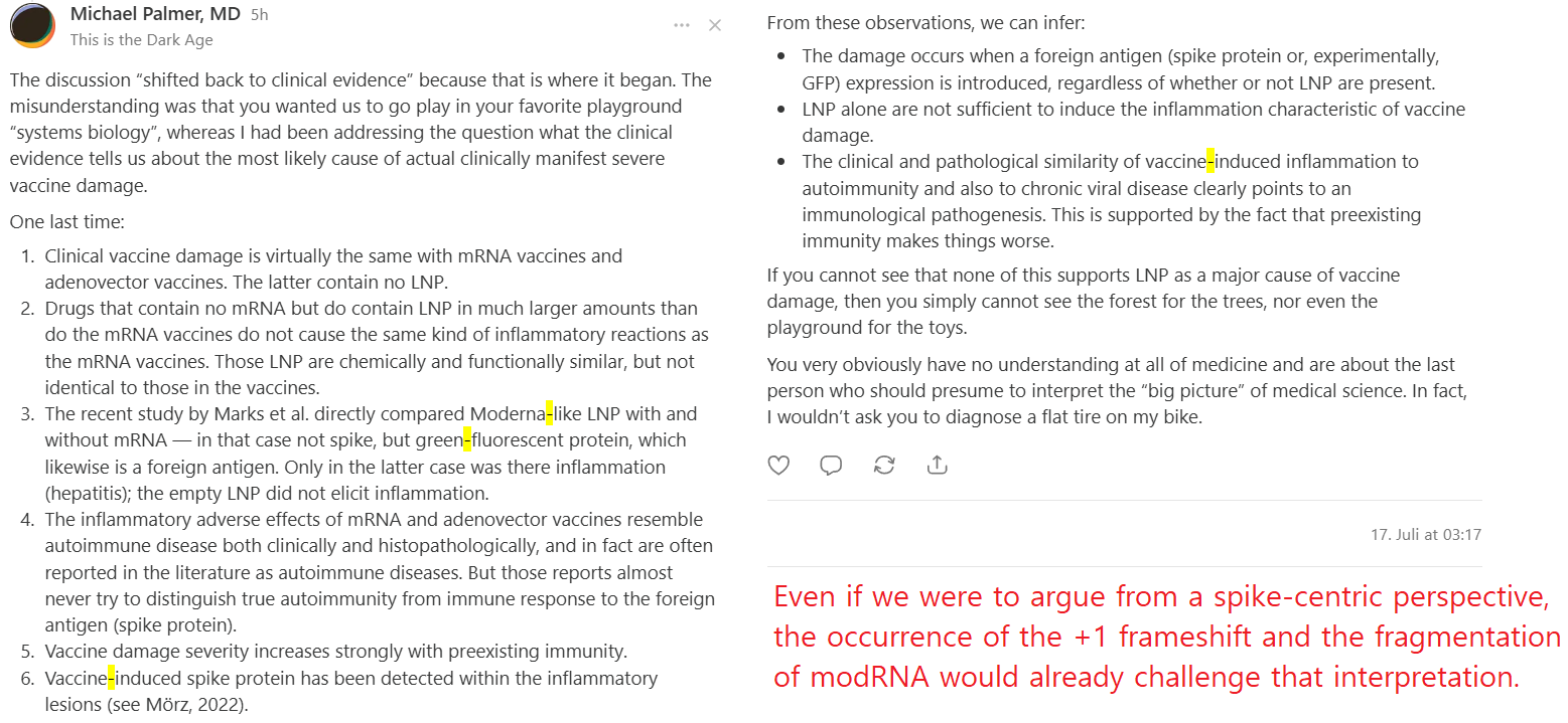
Thanks for this summarizing of the position. However, the reply still does not engage with the central methodological objection, does not answer any of the points that were made, and the closing with ad hominem remarks do nothing to change that.
First, I would like to clarify where our hypothesis actually begins and where Michael Palmer’s argumentation appears to start.
Our framework begins with the earliest biological events following administration of modRNA-LNPs:1
Biodistribution and particle heterogeneity:
This includes differences in lipid species, the presence of empty and partially loaded LNPs within a single formulation, payload-dependent particle properties, and interactions with the protein corona.Physicochemical interactions with cellular membranes:
This includes dielectric and electrostatic properties, membrane interactions, lipid organization, membrane architecture, and effects on intracellular trafficking processes.Downstream signaling pathways and cellular responses:
This includes innate sensing, stress responses, altered signaling networks, and cell-type-specific biological consequences.Systemic consequences:
This includes how local cellular effects may translate into broader tissue-level or organism-level responses.
In contrast, Michael Palmer’s argument appears to begin much further downstream, at what could be considered the final biological outcome: the translated antigen.
He treats this downstream product as the primary and most plausible explanatory mechanism while not sufficiently addressing the upstream processes that precede it, including:
Translational regulation and control mechanisms
Ribosomal organization and regulation
miRNA-mediated regulation, protein folding, and endoplasmic reticulum responses
Potential frame-shifting and alternative translation products
The fundamental methodological difference is therefore not simply a disagreement about the role of the spike protein. It is a disagreement about which level of biological causation should be investigated first.
Our argument is that upstream processes cannot be excluded simply because a downstream product can be detected. Detecting the final product of a biological cascade does not, by itself, identify the initiating event or establish causal primacy.
Therefore, the relevant scientific question is not whether the antigen contributes to some adverse outcomes, but whether the full biological cascade from formulation → cellular interaction → intracellular regulation → antigen expression → immune response has been sufficiently dissected to determine the relative contribution of each component.
What is further important is that the majority of clinical investigations have primarily focused on immune parameters and immune-mediated mechanisms. They have generally not addressed the consequences of different transfected cell populations, such as LSECs, hepatocyctes, Kupffer cells, epithelial cells, monocytes, or other potentially affected cell types.
However, if the biological question concerns causality, restricting the analysis primarily to immune responses is insufficient. Immune activation may represent a downstream amplification mechanism rather than the initial cellular event.
A comprehensive mechanistic evaluation requires investigation of the full sequence of events: which cell types are transfected, how they respond intracellularly, whether altered cellular programs are induced, and how these local effects may subsequently interact with immune pathways.
Without addressing these upstream cellular processes, it remains difficult to determine the relative contribution of antigen expression, innate immune activation, lipid-mediated effects, or other formulation-dependent mechanisms.
1. “Clinical vaccine damage is virtually the same with mRNA vaccines and adenovector vaccines.”
This statement conflates similarity of clinical presentation with similarity of causation and understating mechanisms of action. Different molecular perturbations frequently converge on similar inflammatory endpoints. This is a fundamental property of biological networks and says nothing about whether membrane perturbation contributes to the pathogenic cascade in one platform but not the other.
Additionally, I asked for works demonstrating a comparable broad spectrum of (in a lot of cases idiopathic/idiosyncratic)2,3,4,5,6 adverse events following adenovector vaccinesincluding small-fibre neuropathy, new-onset minimal change disease, cutaneous lymphoma, inflammatory bowel disease, thyroiditis, autoimmune-like hepatitis (which already raises questions about a purely autoimmune-driven interpretation)7, non-Hodgkin lymphoma, anti-GPCR-auto-antibodies (most driven by modRNA-LNP-vaccines and only in very rare cases for Adeno Vector)8 etc.
It is the sum and heterogeneity of these observations, not merely isolated comparable outcomes, that justifies investigating whether a more plausible upstream mechanism exists than simply “It is the spike.”
Additionally, given the well-established importance of bacterial interactions with host membranes in shaping cellular responses, it is surprising that analogous principles of membrane biology are being considered mechanistically negligible in the context of LNP-based RNA delivery. Membrane perturbation is a fundamental biological interface, and its potential consequences should be evaluated experimentally rather than excluded a priori.
Furthermore, the multi-omics findings we discussed point toward disturbances extending beyond a classical immune response. When transcriptomic and proteomic analyses reveal prolonged dysregulation of diverse biological patterns, including cell-cycle-associated pathways persisting for up to 7–10 days (Knabl et al.), and alterations in pathways such as MAPKs identified across different models; including findings persisting for months after a third booster dose in transfected individuals. These observations are compatible with broader cellular stress and/or homeostatic perturbation and therefore warrant mechanistic investigation.
Whether these alterations ultimately translate into clinically relevant pathology is precisely one of the central questions our framework addresses and requires further investigation. Dismissing these observations simply because their clinical significance has not yet been fully established reverses the logic of hypothesis-driven research. Their existence is precisely what motivates mechanistic investigation.
2. “Drugs containing much larger amounts of LNP do not cause the same inflammatory reactions.”
This argument ignores formulation architecture, lipid physicochemistry, biodistribution, administration route, particle composition, payload-dependent particle behavior, and repeated exposure. The example, patisiran, differs substantially from COVID vaccine formulations in all of these respects. I have discussed these distinctions extensively, and I would therefore encourage Michael Palmer to first engage with the underlying reasoning and the relevant literature before returning to lipid quantity as the primary comparison.
This does not prove that LNPs are the dominant clinical driver. It demonstrates only that such comparisons require more than lipid quantity as a variable. This fact does not change because the provided literature is repeatedly ignored. Moreover, several of the papers Michael Palmer cites appear to be interpreted beyond what their experimental design actually demonstrates.
3. “Marks et al. showed that empty LNP did not induce hepatitis.”
To clarify this point again: even if accepted at face value, this demonstrates only that empty LNPs were insufficient to produce hepatitis under the specific experimental conditions studied. It does not demonstrate the absence of membrane perturbation, altered phosphoinositide organization, changes in signaling networks, or cell-type-specific dysfunction.9
Erratum
In my previous assessment, I stated that the study lacked appropriate control formulations to distinguish lipid nanoparticle (LNP)-associated effects from antigen-specific immune responses. This statement was (half) incorrect.
Upon re-examination, I found that I had overlooked the JEDI experimental panel (Fig. 5a–f). In these experiments, the authors explicitly included empty LNPs (Ctrl/eLNP), formulated without mRNA or a non-expressing, as a control. In the separate CAR T-cell experiments (Fig. 5g–l), they additionally used an irrelevant RNA control. My previous criticism regarding the absence of these controls was therefore erroneous and should be withdrawn. The argument will follow this new perspective.
Empty LNP was in fact tested in vivo against RNA.WT and RNA.122T, but only on gross tissue-damage endpoints, hepatic CD8⁺ T-cell infiltration and serum ALT/AST (Fig. 5c,d), within a specific adoptive-transfer model using pre-established, high-avidity antigen-specific T cells. A negative finding on organ-level toxicity and immune infiltration therefore does not rule out such upstream effects on hepatocyte biology, nor does it address LNP-driven pathology that might arise independently of the adoptively transferred T-cell system or only after repeat dosing.
However, the endpoints assessed were relatively coarse. Measurements such as ALT/AST levels and histological analysis of T-cell infiltration detect overt tissue injury and immune cell infiltration but are not designed to capture more subtle, non-lethal effects on membrane organization, phosphoinositide distribution, or intracellular signaling networks. Consequently, negative findings at this level do not exclude the existence of upstream mechanistic effects10,11 ,12
In addition, the Jedi T cell system used for the empty LNP comparison is a monoclonal, TCR transgenic population adoptively transferred at a defined, likely supraphysiological frequency, a tool optimized for sensitively detecting antigen specific killing rather than establishing whether a physiologically primed, polyclonal T cell response following standard vaccination would reproduce the same pattern of hepatocyte sparing. Critically, swapping to the second hepatotoxicity model does not remedy this because the hCD19 CAR T system is equally non physiological, merely substituting a monoclonal, CAR transduced population adoptively transferred at 5 × 10⁶ cells for a monoclonal, TCR transgenic one. Neither model involves an endogenous, polyclonal T cell response elicited by standard vaccination.
Notably, the two features that would most improve external validity, namely a clinically relevant antigen and a naturally primed, polyclonal T cell repertoire, never co occur with a liver damage endpoint anywhere in the dataset. The Spike specific CD8⁺ T cell responses reported elsewhere in the paper (Fig. 3d,e; Fig. 6i,j) arise from endogenous, polyclonal T cells physiologically primed by actual vaccination, yet these Spike experiments were never paired with ALT or AST measurement or hepatic CD8⁺ infiltration. The paper’s hepatotoxicity claim is therefore anchored entirely in artificial, adoptively transferred monoclonal effector systems and a model antigen (GFP) or a therapeutic CAR target (hCD19), and has not been cross validated against a clinically relevant antigen together with a physiologically generated immune response.
Analyses were performed at a single time point (day 5) following a single intravenous (not intramuscular) administration, without repeat dosing or long-term follow-up. Interestingly, Marks et al. note that even intramuscular injection is capable of transfect the liver and a portion ends up in the spleen.
In summary, the experimental design demonstrates that antigen-specific CD8⁺ T cells are sufficient to induce liver injury under the conditions tested. However, it does not establish that these T cells are the sole mediators of the observed pathology. Contributions from innate hepatic cell populations, including LSECs and Kupffer cells were not directly examined and therefore cannot be excluded.
Even if not the case, hepatocyte toxicity would be only one downstream endpoint. Our hypothesis concerns upstream membrane biology and cellular regulation. The interpretation of this experiment appears to be an interesting but highly expansive extrapolation beyond what was actually tested.
Addendum 18.07.2026
I’ll try to summarize, as simply as possible, what this paper actually demonstrated and, equally importantly, what it did not. Because, in the end, it has very little to do with the question of Spike pathogenicity:
They ran a completely separate Spike experiment with a PBS control that doesn’t actually tell you anything about this question. In the relevant experiment, they generated GFP mRNA either without miRNA target sites (WT) or with target sequences for miR-122 or miR-142 inserted into the 3′ UTR, encapsulated in SM-102 LNPs.
After IV injection of the GFP-encoding RNA-LNP (RNA.WT, RNA.122T or RNA.142T) into mice carrying adoptively transferred, monoclonal GFP-specific Jedi T cells, the mice were sacrificed at predetermined time points: 24, 72 and 120 hours (day 1, 3, 5). Liver and spleen were harvested for CD8a immunohistochemistry, serum ALT/AST measurement and flow cytometric quantification of Jedi T cell expansion.
Without assessing bilirubin and other markers of liver injury, this proves nothing more than that the expressed GFP mRNA was a driver for CD8+ monoclonal T cells with a GFP-primed TCR capable of inducing hepatitis.
End of the addendum
There is also a methodological inconsistency that I find difficult to reconcile. On the one hand, Michael Palmer repeatedly asks where the clinically relevant evidence for LNP-mediated mechanisms is. On the other hand, he relies on such highly experimental models to support mechanistic conclusions, despite the fact that such models can generate plausible hypotheses but do not necessarily establish direct clinical relevance. Experimental models are valuable for both sides of this discussion, but their interpretation requires the same standard of caution. Given so, he would also have to accept the sickness behavior study by Korzun et al.13
4. “The pathology resembles autoimmune disease.”
Similar histopathology does not identify the initiating mechanism. Many fundamentally different upstream processes converge on common inflammatory pathways.
Histological similarity is therefore insufficient to distinguish between competing mechanistic models. The relevant question is one of explanatory plausibility, and a model attributing a highly heterogeneous spectrum of outcomes primarily to a single molecular effector requires substantial additional assumptions to maintain explanatory adequacy.
5. “Preexisting immunity increases severity.”
This is entirely compatible with our framework. A stronger immune response in previously sensitized individuals does not exclude additional upstream mechanisms contributing to disease initiation or amplification.
6. “Spike protein has been detected within lesions.”
This is perhaps the most striking example of correlation being mistaken for causal primacy.14
Moerz et al. provide evidence that vaccine-derived spike protein can be detected within encephalitic lesions and interpret this as supporting a causal role of gene-based vaccination. However, the study does not establish that spike protein alone represents the initiating or dominant pathogenic mechanism. Detection of a downstream translated antigen demonstrates biological activity of the vaccine platform but does not resolve the relative contribution of upstream processes, including formulation-dependent biodistribution, cellular uptake, intracellular regulation, or innate cellular responses.
Between LNP administration and spike detection lie multiple biological steps, including partial protonation of i-Lipids, LNP restructuring, forming a proteincorona, membrane interaction, endocytosis, endosomal escape, cytosolic delivery, translation, intracellular trafficking, antigen presentation, immune activation, and extensive cellular cross-talk.
Detecting spike at the end of this sequence demonstrates antigen presence after successful translation. It does not establish that spike is the primary pathogenic driver after vaccination. The presence of a downstream product cannot, by itself, identify the upstream initiating event.
This is precisely why mechanistic biology exists.
7. Finally, the continued reframíng of our review as though its purpose were to prove that LNPs are already established as the dominant clinical cause of vaccine injury
It is not working that way. Our review proposes a mechanistic framework that generates experimentally testable predictions. Refuting such a framework requires engaging with the proposed mechanism itself, not repeatedly returning to the statement that clinical evidence currently favors an immune-mediated interpretation, which remains an interpretation rather than an established mechanistic conclusion.
Throughout this discussion, I have repeatedly raised the same methodological objection: the explanatory power of a spike-only model is inherently constrained by the biology of a single molecular effector. Explaining the remarkable heterogeneity of reported biological responses therefore requires an increasing number of additional assumptions, reducing mechanistic parsimony. (Remarkably, that argument has never actually been addressed.)
Instead, the discussion repeatedly returns to clinical observations while leaving the mechanistic objection unanswered. Personal remarks about my understanding of medicine or whether I could diagnose a flat tire do not alter that reality.
Furthermore, this claim is difficult to reconcile with Mori et al.15 The authors explicitly report that empty LNPs impaired cardiac contractility and induced inflammatory cell infiltration. They conclude that cellular responses to lipid components are among the factors contributing to myocarditis development. They further demonstrate that EGFP mRNA-LNPs also reduce cardiac function, leading them to conclude that spike protein expression is not essential for mRNA vaccine-associated myocarditis.
This appears difficult to reconcile with the repeated assertion that this pathology fundamentally depends on expression of a foreign antigen. If one disagrees with the authors’ interpretation, then please explain why, rather than citing the paper as though it supported the position.
The mouse model has obvious limitations, but these limitations do not invalidate the mechanistic question being investigated.
8. Conclusion
The central issue is not whether our hypothesis is already proven correct, nor whether LNP-mediated mechanisms represent the dominant explanation for all vaccine-associated adverse events. That is not the claim we are making.
The relevant question is whether the hypothesis is biologically plausible, mechanistically coherent, and sufficiently supported by existing evidence to justify rigorous testing and potential falsification.
Our argument is that the hypothesis should not be dismissed simply because alternative explanations, such as antigen-specific immune mechanisms, also exist. Competing mechanisms can coexist, and identifying the relative contribution of each requires appropriately designed experiments.
Importantly, we have not argued that LNP-mediated mechanisms are established clinical drivers. We have argued that they represent a scientifically plausible mechanistic framework that generates testable predictions.
Therefore, the appropriate scientific approach is not to assume our hypothesis is correct, but to actively attempt to falsify it using experiments specifically designed to distinguish payload-dependent effects from LNP-mediated effects.
This question becomes increasingly relevant as substantial efforts are underway to expand and optimize LNP-based RNA technologies. Many current development strategies aim not only to improve delivery efficiency and targeting but also to minimize unintended inflammatory responses. A detailed understanding of the biological interactions of LNP formulations is therefore essential for understanding possible off-target effects.
It is also worth noting that our hypothesis was not introduced as an unsupported speculation in isolation. It was developed within a peer-reviewed scientific context and considered sufficiently plausible and mechanistically grounded to undergo evaluation in a high-selectivity journal. This does not establish that the hypothesis is correct, but it does indicate that the proposed framework is scientifically legitimate and warrants experimental investigation rather than dismissal.
Therefore, I think the most productive conclusion would be:
Can we agree that additional research is both justified and necessary to rigorously test this hypothesis, especially as more LNP-based therapies and vaccines continue to be developed?
However, I wish Michael Palmer the best for further research.
Seger, F., Gutschi, L. M., & Seneff, S. Lipid nanoparticles as active biointerfaces: From membrane interaction to systemic dysregulation. Acta Pharmaceutica Sinica B., 2026 S2211383526004235. doi: 10.1016/j.apsb.2026.07.001
mRNA-1273: Idiopathic thrombocytopenic purpura: case report. Reactions Weekly. 2021;1871(1):264. doi: 10.1007/s40278-021-01783-x. Epub 2021 Sep 4. PMCID: PMC8417645.
Kounis NG, Koniari I, Mplani V, Plotas P, Tsigkas G. First Identified Case of Fatal Fulminant Eosinophilic Myocarditis Following the Initial Dose of the Pfizer-BioNTech mRNA COVID-19 Vaccine (BNT162b2, Comirnaty): an Extremely Rare Idiosyncratic Necrotizing Hypersensitivity Reaction Different to Hypersensitivity or Drug-Induced Myocarditis. J Clin Immunol. 2022 May;42(4):736-737. doi: 10.1007/s10875-022-01228-2. Epub 2022 Feb 17. PMID: 35178640; PMCID: PMC8852986.
Hébert M, Couture S, Schmit I. Bilateral Panuveitis with Occlusive Vasculitis following Coronavirus Disease 2019 Vaccination. Ocul Immunol Inflamm. 2023;31(3):660-664. doi:10.1080/09273948.2022.2042325
Lim XR, Leung BP, Ng CYL, Tan JWL, Chan GYL, Loh CM, Tan GLX, Goh VHH, Wong LT, Chua CR, Tan SC, Lee SSM, Howe HS, Thong BYH, Leong KP. Pseudo-Anaphylactic Reactions to Pfizer BNT162b2 Vaccine: Report of 3 Cases of Anaphylaxis Post Pfizer BNT162b2 Vaccination. Vaccines (Basel). 2021 Aug 31;9(9):974. doi: 10.3390/vaccines9090974. PMID: 34579211; PMCID: PMC8471482.
Buján Bonino C, Moreiras Arias N, López-Pardo Rico M, Pita da Veiga Seijo G, Rosón López E, Suárez Peñaranda JM, Sánchez-Aguilar Rojas D. Atypical erythema multiforme related to BNT162b2 (Pfizer-BioNTech) COVID-19 vaccine. Int J Dermatol. 2021 Nov;60(11):e466-e467. doi: 10.1111/ijd.15894. Epub 2021 Sep 2. PMID: 34473839; PMCID: PMC8652986.
Nyein CM, Liew ZHS, Leow WQ, Yeong PSJ, Ho GH. Severe de novo liver injury after Moderna vaccination - not always autoimmune hepatitis. J Hepatol. 2022 Aug;77(2):556-558. doi: 10.1016/j.jhep.2022.03.041. Epub 2022 Apr 18. PMID: 35439566; PMCID: PMC9051901.
Mantovani M, Bellavite P, Fazio S, Di Fede G, Tomasi M, Belli D, Zanolin E. Autoantibodies Targeting G-Protein-Coupled Receptors and RAS-Related Molecules in Post-Acute COVID Vaccination Syndrome: A Retrospective Case Series Study. Biomedicines. 2024 Dec 15;12(12):2852. doi: 10.3390/biomedicines12122852. PMID: 39767757; PMCID: PMC11673082.
Marks A, Siu S, Bianchini F, et al. mRNA vaccine immunity is enhanced by hepatocyte detargeting and not dependent on dendritic cell expression. Nat Biotechnol., 2026. doi:10.1038/s41587-026-03099-z
Malhi H, Gores GJ. Cellular and molecular mechanisms of liver injury. Gastroenterology. 2008 May;134(6):1641-54. doi: 10.1053/j.gastro.2008.03.002. PMID: 18471544; PMCID: PMC2553363.
Malaguarnera G, Cataudella E, Giordano M, Nunnari G, Chisari G, Malaguarnera M. Toxic hepatitis in occupational exposure to solvents. World J Gastroenterol. 2012 Jun 14;18(22):2756-66. doi: 10.3748/wjg.v18.i22.2756. PMID: 22719183; PMCID: PMC3374978.
Chen CL, Lin YC. Autophagy Dysregulation in Metabolic Associated Fatty Liver Disease: A New Therapeutic Target. Int J Mol Sci. 2022 Sep 2;23(17):10055. doi: 10.3390/ijms231710055. PMID: 36077452; PMCID: PMC9456355.
Korzun T, Moses AS, Jozic A, Grigoriev V, Newton S, Kim J, Diba P, Sattler A, Levasseur PR, Le N, Singh P, Sharma KS, Goo YT, Mamnoon B, Raitmayr C, Mesquita Souza AP, Taratula OR, Sahay G, Taratula O, Marks DL. Lipid Nanoparticles Elicit Reactogenicity and Sickness Behavior in Mice Via Toll-Like Receptor 4 and Myeloid Differentiation Protein 88 Axis. ACS Nano. 2024 Sep 10;18(36):24842-24859. doi: 10.1021/acsnano.4c05088. Epub 2024 Aug 26. PMID: 39186628; PMCID: PMC11916992.
Mörz M. A Case Report: Multifocal Necrotizing Encephalitis and Myocarditis after BNT162b2 mRNA Vaccination against COVID-19. Vaccines (Basel). 2022;10(10):1651. Published 2022 Oct 1. doi:10.3390/vaccines10101651
Mori G, Yamamoto M, Ishikawa K, Tamashiro H, Suzuki H, Mizuno S, Nakada K, Kawaguchi A. Mitochondrial vulnerability underlies myocarditis from COVID-19 mRNA vaccine. Nat Commun. 2026 Apr 1;17(1):4716. doi: 10.1038/s41467-026-71295-1. PMID: 41922346; PMCID: PMC13212923.